File:Nipah.jpg
Nipah.jpg (700 × 527 pixels, file size: 82 KB, MIME type: image/jpeg)
Captions
Captions
Summary
edit| DescriptionNipah.jpg |
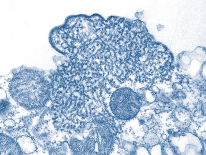
This transmission electron micrograph (TEM) depicted a number of Nipah virus virions that had been isolated from a patient's cerebrospinal fluid (CSF) specimen. Nipah virus is a member of the family Paramyxoviridae, and is related, but not identical, to Hendra virus. Nipah virus was initially isolated in 1999 upon examining samples from an outbreak of encephalitis and respiratory illness among adult men in Malaysia and Singapore. Hendra virus, formerly called equine morbillivirus, is also a member of the family Paramyxoviridae. The virus was first isolated in 1994 from specimens obtained during an outbreak of respiratory and neurologic disease in horses and humans in Hendra, a suburb of Brisbane, Australia. |
| Date | |
| Source | [1] (CDC) |
| Author | CDC/ C. S. Goldsmith, P. E. Rollin |
| Permission (Reusing this file) |
public domain |
Licensing
edit| Public domainPublic domainfalsefalse |
This file is a work of the Centers for Disease Control and Prevention, part of the United States Department of Health and Human Services, taken or made as part of an employee's official duties. As a work of the U.S. federal government, the file is in the public domain.
|
 |
File history
Click on a date/time to view the file as it appeared at that time.
| Date/Time | Thumbnail | Dimensions | User | Comment | |
|---|---|---|---|---|---|
| current | 07:50, 22 May 2008 |  | 700 × 527 (82 KB) | Filip em (talk | contribs) | {{Information |Description=This transmission electron micrograph (TEM) depicted a number of Nipah virus virions that had been isolated from a patient's cerebrospinal fluid (CSF) specimen. Nipah virus is a member of the family Paramyxoviridae, and is rela |
You cannot overwrite this file.
File usage on Commons
The following page uses this file:
File usage on other wikis
The following other wikis use this file:
- Usage on ar.wikipedia.org
- Usage on bg.wikipedia.org
- Usage on ca.wikipedia.org
- Usage on de.wikipedia.org
- Usage on en.wikipedia.org
- Usage on es.wikipedia.org
- Usage on hu.wikipedia.org
- Usage on id.wikipedia.org
- Usage on id.wiktionary.org
- Usage on it.wikipedia.org
- Usage on lv.wikipedia.org
- Usage on nn.wikipedia.org
- Usage on pl.wikipedia.org
- Usage on pt.wikipedia.org
- Usage on ru.wikipedia.org
- Usage on uk.wikipedia.org
- Usage on www.wikidata.org
- Usage on yo.wikipedia.org
- Usage on zh.wikipedia.org
Metadata
This file contains additional information such as Exif metadata which may have been added by the digital camera, scanner, or software program used to create or digitize it. If the file has been modified from its original state, some details such as the timestamp may not fully reflect those of the original file. The timestamp is only as accurate as the clock in the camera, and it may be completely wrong.
| _error | 0 |
|---|
