File:Melas dwi ax.jpg
Size of this preview: 479 × 600 pixels. Other resolutions: 192 × 240 pixels | 539 × 675 pixels.
Original file (539 × 675 pixels, file size: 43 KB, MIME type: image/jpeg)
Captions
Captions
Add a one-line explanation of what this file represents
| Description |
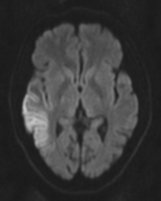
English: "This diffusion-weighted MR image shows cortical ribbon-like high signal consistent with diffusion restriction in a patient with known MELAS and an acute presentation with confusion and focal neurological deficit." (dr Dawes)
Polski: Obraz MR-DWI głowy pacjenta z MELAS, widać pasmo intensywnego sygnału z kory mózgowej, co odpowiada obrazowi klinicznemu (splątanie, ogniskowy deficyt neurologiczny). |
| Date | |
| Source | radpod.org |
| Author | Dr Laughlin Dawes |
| Permission (Reusing this file) |
author kindly mailed me permission to use this and other images on cc-by-3.0 license |
This file is licensed under the Creative Commons Attribution 3.0 Unported license.
- You are free:
- to share – to copy, distribute and transmit the work
- to remix – to adapt the work
- Under the following conditions:
- attribution – You must give appropriate credit, provide a link to the license, and indicate if changes were made. You may do so in any reasonable manner, but not in any way that suggests the licensor endorses you or your use.
File history
Click on a date/time to view the file as it appeared at that time.
| Date/Time | Thumbnail | Dimensions | User | Comment | |
|---|---|---|---|---|---|
| current | 10:01, 15 May 2008 | | 539 × 675 (43 KB) | Filip em (talk | contribs) | {{Information |Description="This diffusion-weighted MR image shows cortical ribbon-like high signal consistent with diffusion restriction in a patient with known MELAS and an acute presentation with confusion and focal neurological deficit. Mitochondrial |
You cannot overwrite this file.
File usage on Commons
There are no pages that use this file.
File usage on other wikis
The following other wikis use this file:
- Usage on ar.wikipedia.org
- Usage on en.wikipedia.org
- Usage on fa.wikipedia.org
- Usage on it.wikipedia.org
- Usage on ja.wikipedia.org
- Usage on pl.wikipedia.org
- Usage on uk.wikipedia.org