File:Eosinophil TEM.jpg
Eosinophil_TEM.jpg (640 × 480 pixels, file size: 69 KB, MIME type: image/jpeg)
File information
Structured data
Captions
Captions
Add a one-line explanation of what this file represents
Summary
edit| DescriptionEosinophil TEM.jpg |
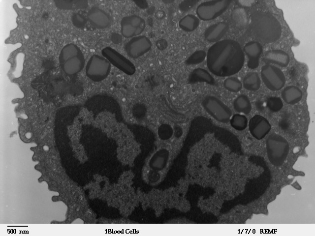
English: Transmission electron microscope image of a thin section cut through a human leukocyte of the type Eosinophil. Eosinophils contain eosinophil granules that are large(0.1-1.0micron) spherical, membrane-bound structures, containing a dense and lamellated crystalloid core. |
| Date | Unknown date |
| Source | http://remf.dartmouth.edu/images/humanBloodCellsTEM/source/1.html |
| Author | Louisa Howard |
| Permission (Reusing this file) |
Public domain as stated by the author, see http://remf.dartmouth.edu/imagesindex.html |
Licensing
edit| Public domainPublic domainfalsefalse |
| I, the copyright holder of this work, release this work into the public domain. This applies worldwide. In some countries this may not be legally possible; if so: I grant anyone the right to use this work for any purpose, without any conditions, unless such conditions are required by law. |
File history
Click on a date/time to view the file as it appeared at that time.
| Date/Time | Thumbnail | Dimensions | User | Comment | |
|---|---|---|---|---|---|
| current | 16:03, 5 November 2009 |  | 640 × 480 (69 KB) | Vojtěch Dostál (talk | contribs) | == Summary == {{Information |Description={{en|1=Transmission electron microscope image of a thin section cut through a human leukocyte of the type Eosinophil. Eosinophils contain eosinophil granules that are large(0.1-1.0micron) spherical, membrane-bound |
You cannot overwrite this file.
File usage on Commons
The following page uses this file:
File usage on other wikis
The following other wikis use this file:
- Usage on cs.wikipedia.org
- Usage on ka.wikipedia.org
- Usage on uk.wikipedia.org
Metadata
This file contains additional information such as Exif metadata which may have been added by the digital camera, scanner, or software program used to create or digitize it. If the file has been modified from its original state, some details such as the timestamp may not fully reflect those of the original file. The timestamp is only as accurate as the clock in the camera, and it may be completely wrong.
| _error | 0 |
|---|
